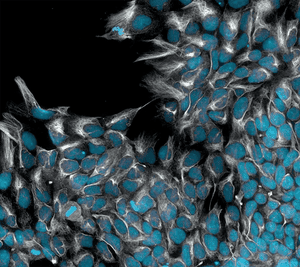
Induced pluripotent stem cells undergoing directed differentiation into early ectoderm. The blue nuclei signify the presence of the Pax6 gene, while the intricate white filaments outline Nestin.
Photograph: Desi Veleva/AIBN
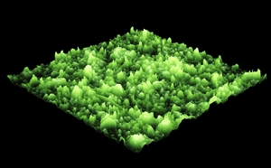
Sea anemone or metal organic framework? Hint: probably best this doesn’t go in the ocean.
Photograph: Silvia Chowdhury/AIBN
Dendrites never looked so magical. What looks like an enchanted forest is in fact a zinc electrode after cycling in an aqueous battery.
Photograph: Yiqing Wang/AIBN
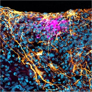
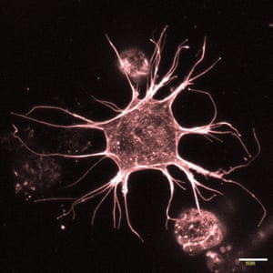
Brain organoids are tiny, synthetic representations of real human brains. This section of a human brain organoid shows the star-shaped glial cells known as astrocytes, aged 140 days.
Photograph: Bahaa Al-mhanawi/AIBN
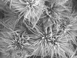
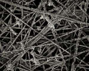
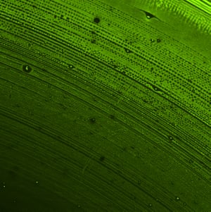
This frosty-looking piece of nanoscience is actually carbon paper substrate that is supporting gold deposition for advanced electrocatalyst development.
Photograph: Asep Nugraha/AIBN
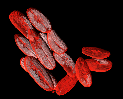
Most people would probably never guess what these are in a million years (it’s a 3D scan of some finger limes).
Photograph: Nicole Atcheson/AIBN
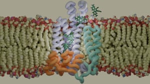
Here, a molecular dynamics simulator has been used to represent a QacA multidrug efflux pump transporting ethidium bromide across the cell membrane in methicillin-resistant staphylococcus aureus.
Illustration: Patrick Sutton/AIBN
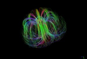
A diffusion MRI tractography was used to map a developing Burderkin plum – specifically the fibrous structures that supply various nutrients.
Photograph: Nyoman Kurniawan/AIBN
Not a palm leaf or the inside of a bottle, but a fluorescein isothiocyanate lipids vacuum-dried down to form a multilayer and imaged on a 10x fluorescence microscope.
Photograph: Aidan Thiele/AIBN
An osteocyte is the most abundant cell in mature bone. Here’s one in a 3D biomimetic hydrogel system.
Photograph: Shiva Muthuswamy/AIBN
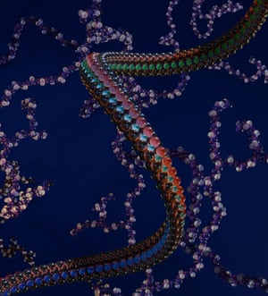
A showcase of digital PolyNIPAM and polyethyleneimine models, created from real polymer simulations via the PolyConstruct platform.
Illustration: Ada Quinn/AIBN
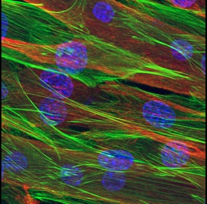
AIBN researchers aren’t typically bodybuilders, but nobody beats them when it comes to growing muscles. Pictured here is myotube grown in a dish using muscle stem cells.
Photograph: Melinder Gill/AIBN
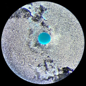
Like distant exoplanet of an unknown world, this image unveils the incredible regenerative potential of dental pulp stem cells. The vibrant blue sphere specifically illuminates the cartilage-like matrix these cells have formed, confirming successful chondrogenesis.
Photograph: Desi Veleva/AIBN